Raik Jar
Abstract
This paper provides an introduction to the new scientific method of Ion-Beam Codicology, IBC, which has been developed by the author at the University of Oxford for the non-sampling physical analysis of papers, inks, and pigments while maintaining the integrity of the historical manuscript. Also discussed is the development of the first dedicated laboratory for the scientific analysis of manuscripts. Furthermore, the paper introduces a variant of IBC in which samples prepared by the conservator or taken from manuscripts are analysed quantitatively with high spatial resolution, of the order of one micron (one millionth of the metre). The aim of the present study is to demonstrate how IBC can be utilised for determining some manuscript characteristics that cannot be obtained otherwise in order to carry out effective scientific research in both conservation and the history of the book. The capabilities of this novel scientific method include: (1) determining the elemental compositions of papers, inks, and pigments; (2) characterising the interaction of ink and surface treatments with paper; (3) determining the location and distribution of impurities; and (4) measuring ink and pigment grain sizes.
It is pointed out in this paper that IBC employs a host of micro-analytical techniques based on using scanning focused beams of energetic ions, which are in the present case the subatomic particles, protons. The ion-beams are utilised for generating instant characteristic radiation signals from a small specific location within a page of a manuscript. Analysis of the signals produces results in the form of numerical data or multi- dimensional maps which can be used for studying the fabrication of manuscripts, such as the composition of ink of a full stop within the text, and also the relevant environmental and conservation effects. The employed scientific techniques are well established: Proton Induced X-Ray Emission (PIXE), and Rutherford Back-Scattering (RBS).
KEY WORDS Scientific conservation, codicology, physical bibliography, Ion-Beam Codicology, Islamic heritage, paper, ink, pigment, material science, manuscript history or the book, writing analysis, scientific analysis, micro-analysis, imaging, applied physics, applied nuclear physics, accelerator, dating, Ion-Beam Analysis, archaeometry, IBC, , PIXE, SPM, x-ray, proton, microbeam, nuclear microscopy.
Introduction
It is not the aim of this paper to elaborate on the universally appreciated cultural significance of Islamic manuscripts and of their conservation requirements. However, what is important to point out is that we know very little about the science of conserving Islamic manuscripts and of studying Islamic papers, inks, and pigments, these limitations are so significant that guardians of the Islamic cultural heritage are often found to be ill equipped to circumvent rising levels of environmental and cultural challenges. Whilst there is presently growing realisation that science should be adopted in this field, it is my thesis that advanced scientific methodologies should be developed for specific tasks and incorporated in the planning and implementation of policies and in developing conservation research. There is now ample evidence to the effect that many past conservation treatments of manuscripts and printed documents have not been particularly successful. This calls for a re-assessment of the traditional approach to conservation. A more advanced approach should in my view be based on a fuller understanding of the materials in question and their interactions, their deterioration characteristics, and the short and long term effects of conservation treatments. Whilst observation and low technology can offer many possibilities in this field,1 they will stop short of facilitating the investigation of the much needed microscopic details which are crucial in this field. Some of these details could be measured using some existing techniques, such as electron microscopy; however, there is a lack of a more comprehensive approach involving dedicated scientific methodologies designed specifically to answer outstanding conservation questions.
It is in response to the above limitations that I have embarked on the development which is reported in this paper. The aim being to endeavour, to devise the first dedicated methodologies and associated laboratory in order to reveal as much as possible about the microscopic characteristics of papers, inks, and pigments within the context of codicology and conservation of Islamic manuscripts. A testament to this commitment is the subject matter of this paper which traces the development of Ion-Beam Codicology, IBC, and the University of Oxford laboratory for the scientific analysis of manuscripts. It is through such an undertaking and future collaborations that I aim to cultivate new trends in research and education within the context of cultural heritage.
It is also relevant to point out that it would be rather inappropriate to introduce this new development without acknowledging the role of many past and present scholars whose humanist approach of observation and study of Islamic texts has contributed so much to our current knowledge in the field2. It is also timely to indicate that there is presently a growing consensus to the effect that contemporary humanistic studies should benefit from complementary scientific details. This could be so not only for confirming the findings of traditional codicology, but also for creating new insights and for acting as a catalyst for more effective traditional studies3. However, although the subject of manuscript conservation would in the long term benefit from learning the history and make-up of Islamic manuscripts, its adoption of modern science, in line with other branches of conservation, is more of an urgent matter4.
Developing Ion-Beam Codicology, IBC5, and the University of Oxford laboratory for the scientific analysis of manuscripts has been a challenging task combining for the first time scientific scholarship with aspects of conservation, the history of the book, and Islamic heritage. This paper reveals that this project has now resulted in the development of highly refined scientific methodologies which can be applied under a controlled safety procedure. Furthermore, it is reported that the extent of the development is well beyond that of other laboratories with interest in the analysis of manuscripts and other historical artefacts. It is also worthwhile to point out that PIXE, which is one of the techniques of Ion-Beam Codicology, has already been applied successfully to European materials, such as the Guthenburg Bible and Greek and Latin manuscripts6.
The Oxford laboratory employs a host of accelerator-based analytical techniques including PIXE, in order to characterise papers, inks and pigments effectively without any chemistry involvement. The main development is to carry out the measurements on full manuscripts without sampling. However, a variant for this development is also available whereby samples can be studied with high spatial resolution. The two systems can measure the elemental compositions at high sensitivity and perform novel two- and three-dimensional imaging of the elemental distributions. However I would like to stress that although this combination of advanced scientific methodologies was developed specifically for answering many significant questions in codicology and conservation, the outcome would not make the traditional methods which are at the disposal of the conservator redundant. Such methods, e. g. optical spectroscopy, can in many cases be adequate on their own for providing suitable answers. In addition, they can compliment IBC by providing preliminary data.
Presented below are details of some background issues in conservation as well as aspects of the scientific approach and methodologies and laboratory design. Also given are some examples of typical IBC scientific results.
Relevance to conservation
It is possible for me to summarise the considerations of the manuscript conservator in terms of the intrinsic and external effects, microscopic and macroscopic, which are exerted on the manuscript throughout its lifetime. The former includes aspects of the natural decay and also the interactions between the different components used in fabricating the manuscript, which in some cases could develop with time into harmful effects. The latter group of effects includes those due to the environment, conservation, and handling. Science can be beneficial for understanding many of these effects and also for developing conservation procedure. For example, it is now quite common to refer to the pH of paper and to carry out a relevant de-acidification procedure. However, I would like to point out that in order for the scientific principles to be effective, it is necessary to develop an understanding of their primary and secondary effects. This, in many cases, can only be achieved using microscopic means. However, although the conventional paper fibre tests and relevant electron microscopy applications can give some supporting information, we are still far from answering such a key question as, for example, de-acidification induced possible incorporation of surface contaminants within the body of the paper and the characteristics of their long term effect. In my view, addressing such issues is important for the development of Islamic manuscript conservation beyond its present state. This can only be achieved if dedicated scientific innovations, such as IBC, are adopted in researching many of the elusive issues faced by the conservator.
In addition to its proposed direct application in developing the conservation process, IBC will also provide the conservator with a unique opportunity to learn about the fabrication of the manuscript and the environmental effects. This is possible because IBC can measure elemental compositions with high sensitivity and also generate three-dimensional elemental distribution maps. In real terms this translates into such an imaginative outcome as characterising a layer of pigment and underlying paper without any chemical means. In this case IBC can identify the following:
- Types and numbers of atoms present in the pigment and paper at high sensitivity in order to determine compositions.
- Grain size of the pigment, nature of the carrier, and thickness of the layer.
- Presence of a surface treatment layer on the paper, plus layer composition, uniformity, and thickness.
- Presence of surface contamination.
- Diameter and density of fibres present in the paper and the elemental distributions within individual fibres.
What is also unique about IBC is that many of these properties can be determined simultaneously, providing an invaluable correlation between several properties of the same object. In effect, this means that we now have unique multi-analytical techniques, within a single scientific facility. This is very attractive indeed from the point of view of conservation where minimum handling of manuscripts is required, although it is not guaranteed that all the above object characteristics can be determined in every study.
It is also appropriate to point out that beyond its relevance to the conservation process development, IBC has the potential of generating universal databases for distinguishing between papers, inks, and pigments. When such information is applied within a historical context to an adequate mass of material, it should be possible to generate unique scientific means for the classification and attribution of Islamic manuscripts.
Scientific Methodologies
In the case of the present work the underlying science of IBC is the controlled short exposure of, for example, a very small part of a scribed letter to a sub-millimetre in diameter focused beam of protons. The proton-beam, known also as ion-beam, consists of a large number of mono-energetic protons travelling in the same direction. The exposure results in instant emission of characteristic radiation revealing microscopic details of the scribe ink and underlying paper. If, for example, the emitted x-rays are detected using the technique of Proton Induced X-ray Emission, PIXE, then the elemental compositions, and not the chemical make-up, can be determined accurately with high sensitivity. Furthermore, it should be noted that in the case of manuscript analysis, PIXE is not a surface technique because papers and inks are transparent to the protons. This is an advantage in comparison with other techniques since the PIXE data in this particular situation are in fact representative of the material bulk. Furthermore, when this technique is applied while the narrow proton-beam is scanning over a small area, then it is possible to generate elemental maps containing information about the lateral distributions of individual elements that form the paper and ink. This operation can be carried out with high spatial resolution, of the order of one micron (one millionth of the metre), on small samples which are placed under vacuum. However, for full manuscripts, and without sampling, the Oxford laboratory facility of the Scanning External Proton Milliprobe7 can perform the task with spatial resolution of about 200 microns whilst the manuscript is kept under atmospheric pressure. Consequently, the relevant technique is described as External-Beam PIXE.
It is also important to point out that application of the PIXE technique at the Oxford laboratory is carried out simultaneously with others, notably the technique of Rutherford Back-Scattering (RBS). In this case, the instantaneous detection of protons scattered from paper, ink, or pigment is carried out during the proton beam exposure. This technique is particularly sensitive to surface layers; and within the present context it is being used for the first time for determining thickness of pigments and ink layers within manuscripts. Another novel application of RBS which I have developed is that of external beam three-dimensional mapping for the investigation of below the surface details of manuscript materials. Application of this technique is given below:
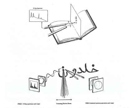
Fig. 1. Sketches of the experimental arrangement used for analysing the ink used in the dot of the letter 'nūn' within a manuscript. In the lop sketch, a stationary beam of protons is used to generate a PIXE x-ray spectrum from the ink. The spectrum contains characteristic lines representing the elements that constitute the ink. According to Ion-Beam Codicology the bottom sketch represents the application of a scanning focused beam of protons in order to generate a spectrum and elemental distribution maps using the PIXE technique, left. In addition, it is also used to generate three-dimensional maps through the RBS technique of the scattering protons, right.
In addition, typical PIXE and RBS spectra from written materials are presented as:
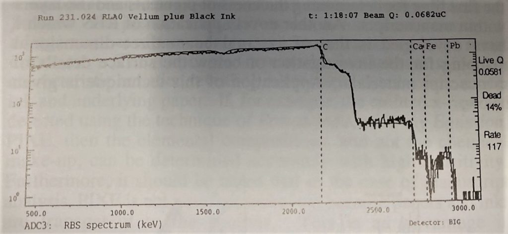
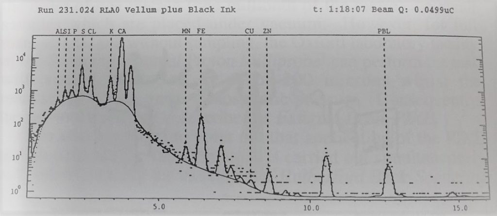
Fig. 2 Spectra generated from a location on a page of a 19th-century book that contains printing black ink on vellum. The top spectrum used the scattered protons of the RBS technique. The spectrum exhibits edges which correspond to the chemical elements present in the ink and vellum, with the leading edge due to the chemical element lead, Pb, present within the printing ink. This edge contains also information about the absolute thickness, of the ink layer. The bottom PIXE x-ray spectrum contains details of the elements that constitute the ink and vellum. Absolute concentrations of the elements including lead and trace elements, can be determined by analysing this spectrum. The two spectra were obtained simultaneously.
Laboratory design
Development of the University of Oxford laboratory was implemented by taking into account both the scientific and conservation safety requirement. In addition, the development programme is an on-going process aimed at future improvements as further operational experience is gained.
Sale handling of the manuscripts is an integral part of the laboratory design brief; this is facilitated by a new manuscript cradle which we developed specifically for the laboratory. This cradle is mobile and can provide the necessary support for manuscripts of different dimensions so that sub-millimetre locations within a given page of a manuscript are selected for analysis. The system is illustrated in Fig. 3 which contains photographs of the cradle and of the manuscript during analysis.
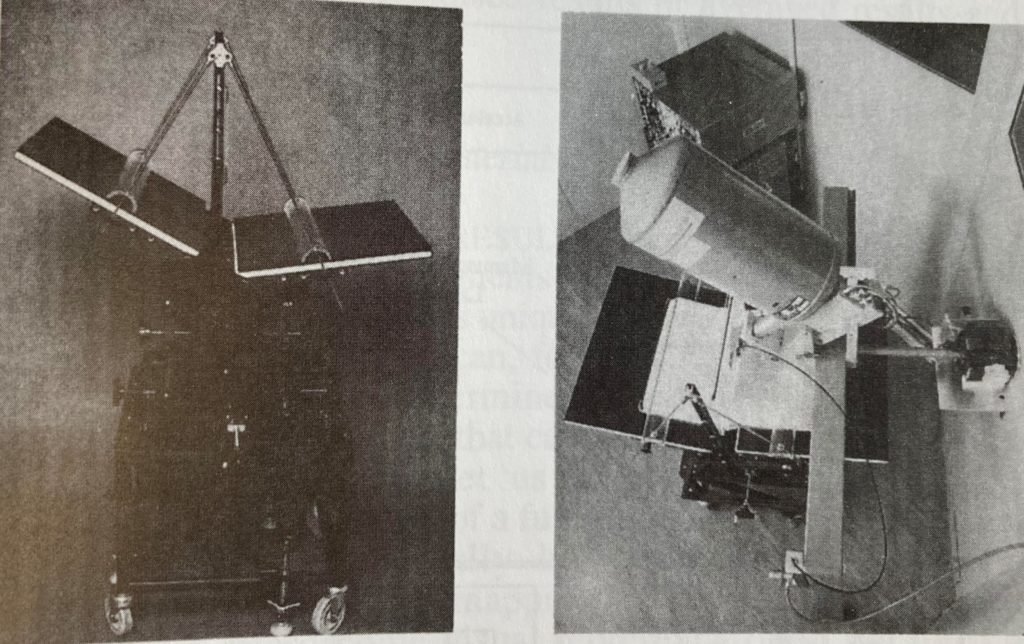
The photograph at the right is a view of the arrangement within the laboratory. The proton beam is directed from the right through the narrow evacuated tube which is terminated by a nozzle for extracting the protons to a small chamber containing helium gas at atmospheric pressure. The manuscript page is kept close to this chamber during analysis and the generated x-rays are detected by the detector which is mounted on top of the supporting frame.
It should be noted that actual analysis is carried out in a special laboratory space defined by partitioning walls and two doors in order to maintain the necessary ambience for handling manuscripts. For safety, the system is computer controlled so that analysis can only proceed when specified conditions are met, such as the closure of doors. Additional safety is provided by pre-setting of the proton beam exposure and on-demand pulsing of the proton beam. These features will ensure minimum exposure conditions as defined by the safety protocol established during numerous tests on samples of Islamic papers, inks, and pigments. The safety protocol also dictates that additional safety tests will have to be incorporated in any forthcoming manuscript analysis projects.
To appreciate the scale of our operation I refer to Fig. 4:
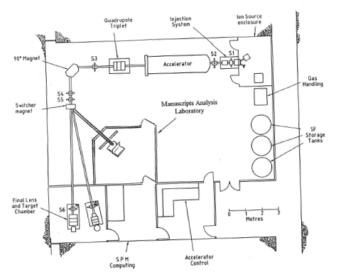
For the technically minded the accelerator is operated by the University of Oxford SPM Unit, and consists of a 1.7MV (mega volt, million volt) Tandem Van de Graaf accelerator producing protons with energy of 3.0MeV (mega electron volt). The protons generated by this accelerator are directed to the manuscripts' analysis laboratory after undergoing magnetic deflection and focusing. The focused proton beam is then extracted within the manuscripts' analysis laboratory through a special thin window material layer at close proximity to the manuscript as shown in Figs. 3 and 4. The instantly emitted radiation generates electronic signals within the radiation detectors. These signals are collected, processed and then stored and displayed on a computer screen, housed in an adjacent room, in the form of spectra and elemental maps. Finally, this manuscript analysis facility can operate while the proton beam is stationary or scanning over the pre-set small area of analysis in order to provide well localised results or averaged results and elemental maps, Fig. 1.
Initial results on historical materials
INTERPRETATION OF IBC RESULTS In order to provide some perspective for the developments which are described above, I remind the reader that IBC is unique in that it is in effect a multi-mode methodology which can, for the first time, and in many cases simultaneously, determine on the atomic scale many characteristics of the object that cannot otherwise be discovered. To simplify this matter let us consider the possibility of characterising the black ink of a full stop not only by determining its composition numerically but also by identifying its components through IBC mapping. The result of the latter possibility is microscopic visual representation in the form of maps appearing in artificial colours to indicate variations in the concentration levels for individual chemical elements present within the area scanned by the proton beam, Fig. 1. For example, if a PIXE elemental distribution map for the element iron is generated then if Fe is actually a constituent of the ink then the full stop would be visualised in the form of localised high concentration within the iron map. In fact we can even image invisible traces of ink if the iron concentration is within the sensitivity limit of the technique. Furthermore, if we simultaneously generate maps for other elements, then we could possibly observe some unique correlation pointing to the true identity of the ink. This can be further established quantitatively by analysing the PIXE spectrum from the ink, which could look like the one given in Fig. 2.
PAPERS The significance of IBC can be demonstrated by a selection of experimental results extracted from a more detailed forthcoming Publication. The first set of results consists of IBC quantative data generated by PIXE analysis of papers in order to prove that the method can provide microscopic means for differentiating between papers. This is particularly interesting because it has been wrongly concluded, by some non-specialists, that because paper is an organic material it is not possible to analyse it using this non-sampling method. What I would like to say at this point is that although it is possible to produce a PIXE eiperimental arrangement which can measure the light elements forming the organic matrix, this is not necessary because the detection sensitivity of PIXE is such that we can measure variation in the concentrations of trace elements present. These trace elements would have originated from the raw material and water used in the fabrication, in order to differentiate between different types of paper. The proof of the matter is that my relevant results, which are given in the table, speak for themselves. These results were obtained under nominal conditions by detecting characteristic x-rays generated by scanning a beam of 3.0MeV protons over paper surface area of . Exposure time of one minute was applied at small proton beam current of 100 pA (pico ampere, one thousandth of a millionth of ampere), in order to detect trace elements at concentrations of parts per million (ppm) in samples of 17th-century Persian paper, 19th-century Turkish calligraphers dyed paper, and modern Archive Text paper of Scottish origin. The minimum detection limits which are given below correspond to the specific experimental conditions. These limits can be improved to few parts per million by increasing the exposure time and/or increasing the x-ray detector solid angle. In addition, in order to perform more detailed paper analysis it is necessary that averaged results of several experiments per sheet are obtained.
Elemental Concentrations in ppm
| Element | Persian | Turkish | Scottish archive | Minimum detectable limit (for 1 minute exposure |
| silicon | 100 | 111 | 580 | 26 |
| sulphur | 502 | 1,088 | 40 | 20 |
| chlorine | 1,223 | 1,042 | 192 | 20 |
| potassium | 1,076 | 708 | 1,739 | 23 |
| calcium | 987 | 2,130 | 10,500 | 30 |
| iron | 198 | 2,907 | 357 | 12 |
| zinc | ـــــــــــــــ | 209 | ـــــــــــــــ | 16 |
It is also important to realise that in addition to the trace element analysis we can now generate elemental distribution maps providing unique information for the conservator. Included in this data are the state of paper surface due to environmental effects and the presence of dyes and other additives, including those due to conservation. The details can be generated by our method of high lateral resolution PLXE and RBS elemental mapping of the paper surface and cross-section, as shown in Fig. 5 (a and b), which includes two sets of two-dimensional elemental maps.
The first set corresponds to the surface of a particular white paper, which exhibits the presence of some heavy metals and an apparent non-uniformity in the elemental distribution in the form of segregated elemental concentrations. The second set of maps was generated by presenting to the proton beam the edge of a 19th-century polished blue paper. In this case one can observe the ends of the fibres which are apparent in the calcium map, and also detect an evidence of surface treatment, at the right-band side of the paper, in the form of enhanced chlorine concentration which is correlated with silicon and aluminium and related to oxygen depletion. It should be also noted that in addition to the major elements, trace element mapping is included in this procedure, and that it is also possible to produce numerical data for the diameters and densities of fibres plus the thickness of surface layers.
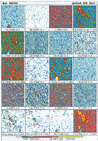
Fig. 5 (a). Ion-Beam Codicology. High resolution elemental mapping of paper. Each frame corresponds to the variation in concentration of a specific chemical element shown as variation in artificial colours with the colour yellow denoting the highest concentration. The results were produced on 19th-century European paper. This is the group of maps (frame size 0.25mm) for the surface of white paper.
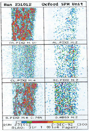
Fig. 5 (b). The group of maps (frame size 0.3mm) for the edge of blue polished paper.
As a secondary development, I have also considered the possibility of complementing IBC results with those based on the arrangement and frequency of chain lines and laid lines. To do this it is possible to use beta-radiography which is a method employed by some scholars to record watermarks, etc. This method employs the radiation emitted from a plate coated with a radioactive substance to generate an image on photographic emulsion after penetrating the paper. However, the technique has three limitations: handling the radioactive source, prolonged exposure over many hours, and limited surface area of the paper that can be irradiated at a time. Following, suggestions by D. Woodward, professor at the University of Wisconsin-Madison, I have now developed a photographic method to record the chain lines and laid lines over the entire manuscript page. This complementary method involves only a few minutes' operation producing remarkable results as shown in Figs. 6 and 7.
Fig. 6. Result of the beta radiography including a record of the chain lines and laid lines within a part of a page from an Islamic manuscript. The image required several hours to exposure.
Fig. 7. Result of the photographic method developed to record the laid lines and chain lines for full pages. The result was produced within minutes on the same manuscript shown in Fig. 6.
INKS AND PIGMENTS Traditionally, ink and pigment analysis using PIXE applied to European materials involves identifying the elemental constituents in the presence of background due to the underlying paper. Although this method on its own has yielded very interesting results indeed, the advent of Ion-Beam Codicology, IBC, which employs scanning focused beams of ions, means that we can now compliment the standard PIXE data with the mapping methodology available at the Oxford laboratory. The other advantage of this development is that instant recognition of the type of inks and pigments can be formulated before any analysis of spectra takes place. In addition, IBC has another unique advantage in that it is also possible to perform three-dimensional mapping in order to characterise ink and pigment layer thickness and possibly to identify the way the scribe applied those layers.
This method of generating two and three-dimensional characterisations of ink or pigment layers within pages of manuscripts while the whole manuscript is supported on its cradle is new in this field and I have given it the title External Beam Multi-Dimensional Analysis (EBMA). In order to demonstrate the potential of EBMA I would like to refer to a given situation of characterising layers of gold, blue pigment and black ink present on a page of a 17th-century Persian manuscript, Fig. 8.
Fig. 8. Photography a page of a 17th-century Persian manuscript with text in black ink and a border of adjacent gold and blue lines.
In this case, it has been possible to characterise the gold and blue adjacent lines which form the border to the text simultaneously by generating maps of selected elements within a mapping frame size of 2mm square, incorporating both the lines. The results which are given in Fig. 9 (a and b) indicate that the gold line consists of an alloy containing gold, copper, and zinc, which has been applied using a sulpher-containing medium, and which has uniform width throughout its thickness. In addition, the blue line consists of a pigment of smalt blue, Fig. 9 (c). A separate test of the black ink reveals that it contains iron and copper plus concentrations of calcium and sulphur, Fig. 9 (d).
Fig. 9 (a). Ion-Beam Codicology of the gold line within the page of the Persian manuscript, Fig. 8. Elemental distribution maps for gold, copper, sulphur, and zinc. The frame size is 2mm square; for a colour map white denotes the highest concentration. However, for a mono-colour map, black represents the highest elemental concentration, and this applies to all maps in Fig. 9 (a-d).
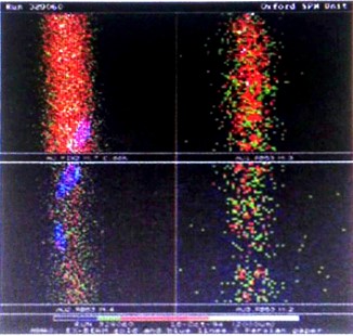
Fig. 9 (b). Ion-Beam Codicology (External Beam Multi-Dimensional Analysis EBMA). Results for the gold line within the page of the Persian manuscript, Fig. 8. Shown are gold distribution at the surface (maps Au.PIX2 and Au1.RBS3) at the interface with the paper (map Au3.RBS3), and an intermediate depth within the gold layer (Au2.RI3S3). Frame size is 2mm square.
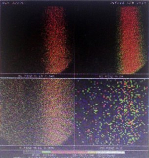
Fig. 9 (c). Ion-Beam Codicology of the blue line within the page of the Persian manuscript, Fig. 8. Elemental distribution maps for aluminum, silicon, cobalt, and potassium. The frame size is 2mm square; the colour white denotes the highest concentration.
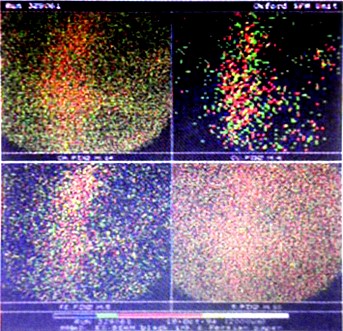
Fig. 9 (d). Ion-Beam Codicology of the black ink within the page of the Persian manuscript, Fig. 8. Elemental distribution maps for calcium, copper, sulphur, and iron. The frame size is 2mm square; the colour white denotes the highest concentration.
These are initial results, and therefore, I shall not at this stage speculate about the nature of the ink till further tests are conducted. However, I would like to indicate that it is also possible using this method to detect the presence of ink which is not visible to the naked eye on the condition that the ink traces are within the detection limits of the analytical technique.
Realities and misconceptions
The introduction of advanced science to conservation is not an easy task, not least because one has to circumvent many misconceptions. This is a key issue which has to be addressed within the broader field of conservation because it is related to many ethical considerations. As far as the present scientific work is concerned, it is first of all important to point out that although IBC is potentially significant it is not claimed that the method will answer all the questions raised now or in the future. It therefore follows that I advocate a more learned approach encompassing the fact that in some cases one has to consider applying more than one technique. It is in recognition of this that I have developed IBC as a multi-techniques method, and introduced the complementary optical method for recording the chain lines and laid lines. Furthermore, it is important to indicate that standard PIXE, on its own, has already proved to be sufficient for characterising inks without the need to determine the chemical make-up directly. In the light of the present work it is obvious that IBC has the potential of performing this more comprehensively. However, if there are some cases when details about the chemical bonding, etc., are required, then I would recommend the application of a complementary technique.
The second important issue is any possible damage to the historical material. This paper gives details of the rare that has been taken in the design of the laboratory and the manuscript cradle, and also in determining safe proton exposure conditions. In the case of the latter, I would like to correct a relevant misconception by some non-specialists who are not familiar with the procedure or with science in general. IBC involves proton exposure of very small locations within a page of a manuscript. What we need to be familiar with in this case is that protons are somewhat similar to light in that they represent a form of energy which if used incorrectly can lead to side effects. For this particular reason I introduced proton dosage control, and developed the equipment so that only very small doses are required for producing the necessary results.
A further element in the safety procedure is that preliminary tests on material standards will precede any dedicated analysis of manuscripts. Furthermore, I would like to state that under the specific Oxford laboratory conditions no radioactivity is introduced, and I observe no side effects on most materials. However, in developing the laboratory procedure I have had to establish safety margins which were developed by using much higher doses of protons on a selection of Islamic materials with the specific purpose of introducing damage. Under these extreme conditions it is possible in some cases to detect slight colouration which could disappear immediately or persist after time8.
Conclusions
The details given in this paper are challenging from the point of view of their intellectual significance for the conservation and study of Islamic manuscripts. They are also challenging for some beliefs that conservation is the domain of conventional technology only. Whilst this can be so in many cases, contemporary requirements indicate that the future of conservation should benefit from science. In fact in such cases as the microscopic details of manuscripts, I can find no other alternative but to develop advanced scientific methodologies. The message of this endeavour is that we should learn as much as possible about the science of manuscripts and the microscopic effects of any proposed conservation treatments.
It is also worthwhile to point out that these aspirations, which are also held by others, cannot flourish in isolation. Therefore, I would like to take this opportunity to urge a positive response to new ideas and to encourage collaboration across the disciplines. After all, it was only through multi-disciplinary efforts and the support of others that Ion-beam Codicology was conceived.
Finally, the present work provides ample evidence to the potential of IBC as a unique multi-techniques method that can be applied within the same laboratory under well controlled conditions. The method lends itself also to complementary efforts involving more traditional methods and it can be applied to determine several unique manuscript characteristics simultaneously and for the first time. Furthermore, the choice provided by the high spatial resolution mapping of historical or prepared samples can be also valuable for determining some of the characteristics of conservation treatment methods. To my knowledge this is the first time that a scientific method and a dedicated laboratory have been developed for the scientific analysis of historical manuscripts. Such an endeavour should at least facilitate advancing our knowledge of the history and conservation of Islamic manuscripts.
I would like to express my gratitude for receiving a grant from the Wellcome Trust, London; which facilitated carrying out a collaborative feasibility study that preceded this work.
The manuscript analysis laboratory is an integral part of the SPM facilities at Oxford. The laboratory technical developments were carried out in collaboration with G. W. Grime, M. Marsh and M. Dawson.
| Source note: This article was published in the following book: The Conservation and Preservation of Islamic Manuscripts, Proceedings of the third conference of Al-Furqān Islamic Heritage Foundation, 18th-19th November 1995 - English version, 1995, Al-Furqān Islamic Heritage Foundation, London, UK, pp. 93-118. |